Introduction:
Oral leukoplakia is one of the most common potentially malignant disorders affecting the oral cavity. It presents as a persistent white patch or plaque inside the mouth that cannot be rubbed off and cannot be clinically or pathologically classified as any other disease. While many cases remain benign, some may progress to oral cancer if left untreated.
Because oral cancer is a major health concern in India, especially among tobacco and gutkha users, early detection and management of oral leukoplakia are crucial. Understanding the causes, symptoms, risk factors, diagnosis, treatment options, and preventive measures can help reduce complications and improve long-term outcomes.
This comprehensive guide covers oral leukoplakia from both modern medical and Ayurvedic perspectives, including epidemiology, prevalence, cancer risk, diagnosis, treatment, diet, lifestyle recommendations, and frequently asked questions.
What Is Oral Leukoplakia?
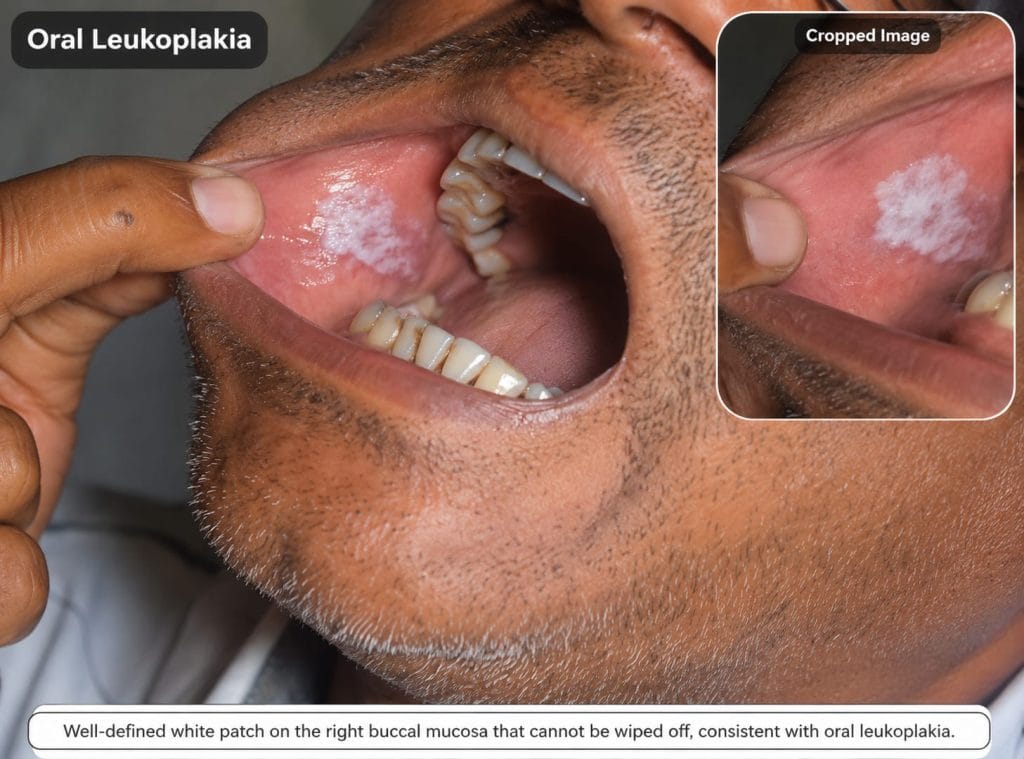
Oral leukoplakia is a predominantly white patch or plaque that develops on the oral mucosa and cannot be removed by scraping. It is considered a potentially malignant disorder because some lesions may transform into oral squamous cell carcinoma over time.
The condition most commonly affects the tongue, inner cheeks, gums, floor of the mouth, and lips.
Key Facts About Oral Leukoplakia:
- Most common potentially malignant disorder of the oral cavity.
- Appears as a persistent white patch that cannot be rubbed off.
- Strongly associated with tobacco, gutkha, smoking, and areca nut chewing.
- Usually painless during early stages.
- Some lesions may become cancerous.
- Early diagnosis significantly improves outcomes.
- Biopsy is often necessary to determine cancer risk.
Epidemiology and Prevalence of Oral Leukoplakia:
Global Epidemiology:
Oral leukoplakia affects approximately 1–4% of the global population. It is one of the most frequently diagnosed oral potentially malignant disorders worldwide.
The prevalence varies according to:
- Tobacco consumption
- Alcohol use
- Betel nut chewing habits
- Socioeconomic factors
- Geographic location
Prevalence in India:
India reports one of the highest rates of oral leukoplakia due to the widespread use of:
- Gutkha
- Pan masala
- Tobacco chewing
- Smoking
- Betel quid
Several studies have reported prevalence rates ranging from 0.7% to 12% in different Indian populations.
Age Distribution:
The condition is most commonly diagnosed between:
- 40 and 70 years of age
Risk increases with prolonged exposure to tobacco and oral irritants.
Gender Distribution:
Historically:
- Men are affected more commonly than women.
- Male-to-female ratio ranges from 2:1 to 3:1.
However, increasing tobacco exposure among women has narrowed this gap.
Public Health Significance:
Oral leukoplakia is considered a significant public health concern because:
- It is often asymptomatic.
- Patients may remain undiagnosed for years.
- It can progress to oral cancer.
- Early screening can detect lesions before malignant transformation occurs.
Causes of Oral Leukoplakia:
Tobacco Use:
Tobacco remains the most important risk factor.
Forms include:
- Cigarette smoking
- Bidi smoking
- Cigar smoking
- Hookah smoking
Smokeless Tobacco and Gutkha:
Smokeless tobacco products greatly increase risk.
Examples:
- Gutkha
- Khaini
- Pan masala
- Zarda
Areca Nut (Betel Nut) Chewing:
Areca nut contains carcinogenic compounds that contribute to mucosal damage and precancerous changes.
Alcohol Consumption:
Heavy alcohol intake acts synergistically with tobacco and increases cancer risk.
Chronic Mechanical Irritation:
Long-standing irritation from:
- Sharp teeth
- Broken fillings
- Ill-fitting dentures
may contribute to lesion formation.
Human Papillomavirus (HPV):
Certain HPV strains have been implicated in oral mucosal lesions.
Nutritional Deficiencies:
Deficiencies of:
- Vitamin A
- Vitamin B Complex
- Iron
- Antioxidants
may increase susceptibility.
Genetic Predisposition:
Genetic and molecular factors may influence the risk of malignant transformation.
Risk Factors for Oral Leukoplakia:
- Tobacco use
- Gutkha consumption
- Pan masala chewing
- Betel nut chewing
- Alcohol use
- Poor oral hygiene
- Nutritional deficiencies
- Chronic irritation
- Increasing age
- Previous oral lesions
Symptoms of Oral Leukoplakia:
Many patients experience no symptoms initially.
Common symptoms include:
- Persistent white patch
- Thickened oral mucosa
- Rough texture
- Wrinkled appearance
- Cracked lesion
- Mild burning sensation
- Oral discomfort
Advanced lesions may cause:
- Pain
- Difficulty chewing
- Difficulty swallowing
- Ulceration
Common Sites of Oral Leukoplakia:
Tongue:
Particularly the lateral borders.
Buccal Mucosa:
Inner lining of the cheeks.
Gums:
Gingival involvement may occur.
Floor of Mouth:
High-risk site for malignant transformation.
Lips and Palate:
Less commonly affected.
Types of Oral Leukoplakia:
Homogeneous Leukoplakia:
Characteristics:
- Uniform white appearance
- Smooth surface
- Flat lesion
- Lower cancer risk
Non-Homogeneous Leukoplakia:
Characteristics:
- Mixed red and white areas
- Nodular surface
- Verrucous appearance
- Higher cancer risk
Proliferative Verrucous Leukoplakia (PVL):
A rare but aggressive variant characterized by:
- Multifocal lesions
- High recurrence rate
- Significant malignant potential
Hairy Leukoplakia vs Oral Leukoplakia:
What Is Hairy Leukoplakia?
Hairy leukoplakia is a white corrugated lesion caused by Epstein-Barr Virus (EBV), usually occurring in immunocompromised individuals.
Causes of Hairy Leukoplakia:
- Epstein-Barr Virus infection
- HIV infection
- Immunosuppression
Clinical Features of Hairy Leukoplakia:
- White corrugated lesion
- Hair-like projections
- Usually on lateral tongue
- Painless
- Non-precancerous
Difference Between Hairy Leukoplakia and Oral Leukoplakia:
| Feature | Hairy Leukoplakia | Oral Leukoplakia |
|---|---|---|
| Cause | EBV infection | Tobacco and irritants |
| Cancer Risk | Usually not precancerous | Potentially malignant |
| Common Site | Lateral tongue | Multiple oral sites |
| Associated Condition | Immunosuppression | Tobacco habits |
| Treatment | Immune restoration | Risk factor elimination and monitoring |
Oral Leukoplakia and Cancer Risk:
Why Is Oral Leukoplakia Considered Precancerous?
Abnormal epithelial changes may gradually progress toward malignancy.
Factors Increasing Malignant Transformation:
- Long-standing lesion
- Non-homogeneous lesion
- Large lesion size
- Female gender
- Tongue involvement
- Floor of mouth involvement
High-Risk Features:
- Red and white lesions
- Nodular lesions
- Verrucous lesions
- Severe dysplasia
Malignant Transformation Rate:
Reported rates vary from 1% to 20% depending on lesion characteristics.
Pathophysiology of Oral Leukoplakia:
Continuous exposure to tobacco and chemical irritants causes:
- Chronic inflammation
- Hyperkeratosis
- Epithelial hyperplasia
- Cellular dysplasia
Progressive genetic mutations may eventually lead to oral cancer.
Diagnosis of Oral Leukoplakia:
Clinical Examination:
Assessment of:
- Size
- Shape
- Color
- Surface texture
Oral Cancer Screening:
Comprehensive oral examination helps identify suspicious lesions.
Medical History:
Assessment of:
- Tobacco use
- Alcohol intake
- Previous lesions
Toluidine Blue Staining:
May assist in identifying high-risk areas.
Brush Cytology:
Used in selected cases.
Biopsy:
Gold standard diagnostic tool.
Histopathological Findings:
Hyperkeratosis:
Increased keratin formation.
Epithelial Hyperplasia:
Increased epithelial thickness.
Mild Dysplasia:
Early cellular abnormalities.
Moderate Dysplasia:
Intermediate-grade abnormalities.
Severe Dysplasia:
Advanced precancerous changes.
Carcinoma In Situ:
Severe epithelial changes without invasion.
Differential Diagnosis of Oral Leukoplakia:
Oral Candidiasis:
Fungal infection that may resemble leukoplakia.
Oral Lichen Planus:
Chronic inflammatory condition.
Frictional Keratosis:
Response to chronic trauma.
White Sponge Nevus:
Genetic oral disorder.
Discoid Lupus Erythematosus:
Autoimmune disease affecting oral mucosa.
Conventional Treatment of Oral Leukoplakia:
Tobacco Cessation:
Most important intervention.
Removal of Local Irritants:
Correction of dental factors.
Surgical Excision:
Recommended for high-risk lesions.
Laser Therapy:
Effective for selected cases.
Cryotherapy:
May be used in specific situations.
Regular Follow-Up:
Essential because recurrence can occur.
Ayurvedic Correlation of Oral Leukoplakia:
Although not directly described in classical texts, oral leukoplakia can be correlated with chronic Mukharoga involving Kapha-Pitta Dushti, Rakta Dushti, and Mamsa Dhatu involvement.
Dosha Involvement:
Kapha Dosha:
Responsible for:
- White plaque formation
- Thickening
- Tissue overgrowth
Pitta Dosha:
Responsible for:
- Inflammation
- Burning sensation
- Malignant tendency
Vata Dosha:
Responsible for:
- Chronicity
- Degeneration
- Irregular morphology
Dushya Involvement:
- Rasa Dhatu
- Rakta Dhatu
- Mamsa Dhatu
Srotas Involvement:
- Rasavaha Srotas
- Raktavaha Srotas
- Mamsavaha Srotas
Ayurvedic Samprapti (Pathogenesis):
Continuous exposure to tobacco, chemical irritants, improper diet, and poor oral hygiene leads to Dosha vitiation, Dhatu Dushti, and chronic oral mucosal changes.
Ayurvedic Treatment of Oral Leukoplakia:
Important Disclaimer: Every persistent white patch should undergo biopsy and evaluation by a qualified dental specialist before initiating Ayurvedic treatment.
Nidana Parivarjana (Elimination of Causes):
- Stop tobacco completely.
- Avoid gutkha.
- Avoid areca nut.
- Limit alcohol.
- Correct dental irritation.
Shodhana Chikitsa:
Virechana Karma:
virechan will Helps eliminate aggravated Pitta and supports Rakta Shodhana.
Nasya Karma:
Supports oral and upper airway health.
Gandusha:
Medicated oil holding helps improve oral hygiene and mucosal health.
Kavala:
Therapeutic gargling helps maintain oral cleanliness.
Shamana Chikitsa:
Individualized Ayurvedic management focuses on:
- Pitta Shamana
- Kapha Shamana
- Rakta Prasadana
- Mucosal healing
Rasayana Chikitsa:
Rasayana therapies aim to:
- Improve tissue repair
- Enhance immunity
- Support cellular health
- Promote long-term wellness
Importance of Long-Term Follow-Up:
Regular oral examination remains essential even when symptoms improve.
Diet for Oral Leukoplakia (Pathya-Apathya):
Recommended Foods (Pathya):
Amla:
Rich source of antioxidants and Vitamin C.
Pomegranate:
Supports mucosal healing.
Carrots:
Rich in beta-carotene.
Green Leafy Vegetables:
Provide protective phytonutrients.
Seasonal Fruits:
Supply vitamins and antioxidants.
Moong Dal:
Light and easy to digest.
Coconut Water:
Supports hydration.
Warm Water:
Promotes digestive health.
Foods to Avoid (Apathya):
Tobacco Products:
Avoid completely.
Gutkha and Pan Masala:
Major risk factors for oral cancer.
Areca Nut:
Associated with precancerous lesions.
Alcohol:
Increases malignant potential.
Excessively Spicy Foods:
May irritate oral mucosa.
Deep-Fried Foods:
Can aggravate inflammation.
Processed Foods:
Contain harmful additives and preservatives.
Carbonated Drinks:
May irritate oral tissues.
Excess Refined Sugar:
May contribute to chronic inflammation.
Very Hot Beverages:
Repeated thermal injury may worsen mucosal damage.
Lifestyle Recommendations for Oral Leukoplakia:
Maintain Oral Hygiene:
Brush twice daily and maintain oral cleanliness.
Regular Dental Checkups:
Early detection improves outcomes.
Avoid Self-Medication:
Seek professional guidance.
Quit Tobacco Completely:
Most important preventive measure.
Stress Management:
Practice yoga and meditation.
Adequate Sleep:
Supports healing and immunity.
Prevention of Oral Leukoplakia:
- Avoid tobacco in all forms.
- Stop gutkha and areca nut use.
- Limit alcohol consumption.
- Maintain good oral hygiene.
- Eat antioxidant-rich foods.
- Undergo regular oral screening.
When Should You See a Doctor?
Seek medical attention if:
- A white patch persists for more than two weeks.
- The lesion enlarges.
- Red areas develop.
- Pain or ulceration occurs.
- Difficulty swallowing develops.
Prognosis of Oral Leukoplakia:
The prognosis depends upon:
- Histopathology
- Lesion type
- Tobacco exposure
- Treatment compliance
Patients who stop tobacco and attend regular follow-ups generally have better outcomes.
Frequently Asked Questions (FAQs)
Is oral leukoplakia cancer?
No. Oral leukoplakia is not cancer but may become cancerous in some cases.
Can oral leukoplakia become cancerous?
Yes. Certain lesions may transform into oral cancer.
Can oral leukoplakia be cured?
Many lesions improve after eliminating risk factors and receiving appropriate treatment.
Is biopsy necessary for oral leukoplakia?
Biopsy is often required to assess cancer risk.
What is the best treatment for oral leukoplakia?
Risk factor elimination, biopsy-based management, and regular follow-up are essential.
Can Ayurveda treat oral leukoplakia?
Ayurveda may support overall oral health and complement conventional care, but biopsy and specialist evaluation should never be delayed.
What foods should be avoided in oral leukoplakia?
Tobacco, gutkha, alcohol, areca nut, excessively spicy foods, and processed foods should be avoided.
What is the difference between hairy leukoplakia and oral leukoplakia?
Hairy leukoplakia is associated with EBV infection and immunosuppression, whereas oral leukoplakia is primarily linked to tobacco exposure and carries a risk of cancer.
Conclusion :
Oral leukoplakia is a potentially malignant oral disorder that should never be ignored. Early diagnosis, elimination of tobacco habits, biopsy of suspicious lesions, regular follow-up, and appropriate treatment are essential to prevent progression to oral cancer. Along with modern medical care, Ayurvedic principles emphasizing Nidana Parivarjana, oral hygiene, proper diet, and lifestyle modifications may help support long-term oral health and overall well-being.
References:
- World Health Organization (WHO)
- National Cancer Institute (NCI)
- American Academy of Oral Medicine
- Burket’s Oral Medicine
- Neville BW, Oral and Maxillofacial Pathology
- PubMed Indexed Articles on Oral Leukoplakia
- Charaka Samhita
- Sushruta Samhita
- Ashtanga Hridaya
Author:
Dr. Shailesh Phalle
Medical Reviewer:
Dr. Shailesh Phalle
Medical Disclaimer:
The information provided in this article is intended for educational and informational purposes only and should not be considered a substitute for professional medical advice, diagnosis, or treatment. Oral leukoplakia is a potentially malignant oral disorder, and any persistent white patch, ulcer, or abnormal lesion in the mouth should be evaluated by a qualified dental surgeon, oral medicine specialist, or healthcare professional.
Ayurvedic treatments discussed in this article are based on classical Ayurvedic principles and clinical experience. Treatment recommendations may vary depending on the individual’s constitution (Prakriti), disease stage, associated medical conditions, and overall health status. Self-diagnosis and self-medication are strongly discouraged.
Patients should not delay or avoid medical evaluation, biopsy, laboratory investigations, or conventional treatment when indicated. Ayurveda may be used as a complementary approach under the supervision of a qualified Ayurvedic physician after appropriate diagnosis and assessment.
The content of this article has been reviewed for medical accuracy; however, outcomes may vary from person to person, and no guarantee of specific treatment results is implied.